


There’s a moment every photographer knows. You’re standing in fading light, the scene in front of you is extraordinary, and you’re excited! Your camera captures something technically correct but – oh no – it’s somehow flat.
The histogram looks fine. The focus is sharp. But the image misses the feeling you had when you pressed the shutter.
That gap – between what you saw and what the camera recorded – lives entirely in your visual system. The camera records light. You experience it. And that experience runs through one of the most complex optical and neurological instruments ever assembled: the human eye.
Most photographers spend thousands on glass to improve the image between the scene and the sensor. But do we think about the visual system between the scene and the brain?
That’s where the real photograph happens.
Two Systems, One Vision
Your eye operates through two distinct muscle systems, and understanding them changes how you think about fatigue, focus, and long-term visual health.
The pointing system. Six muscles surround each eyeball – four recti muscles pulling in the cardinal directions, and two oblique muscles handling rotation. They originate from a fibrous ring around the optic nerve and fire in coordinated pairs to aim your gaze.
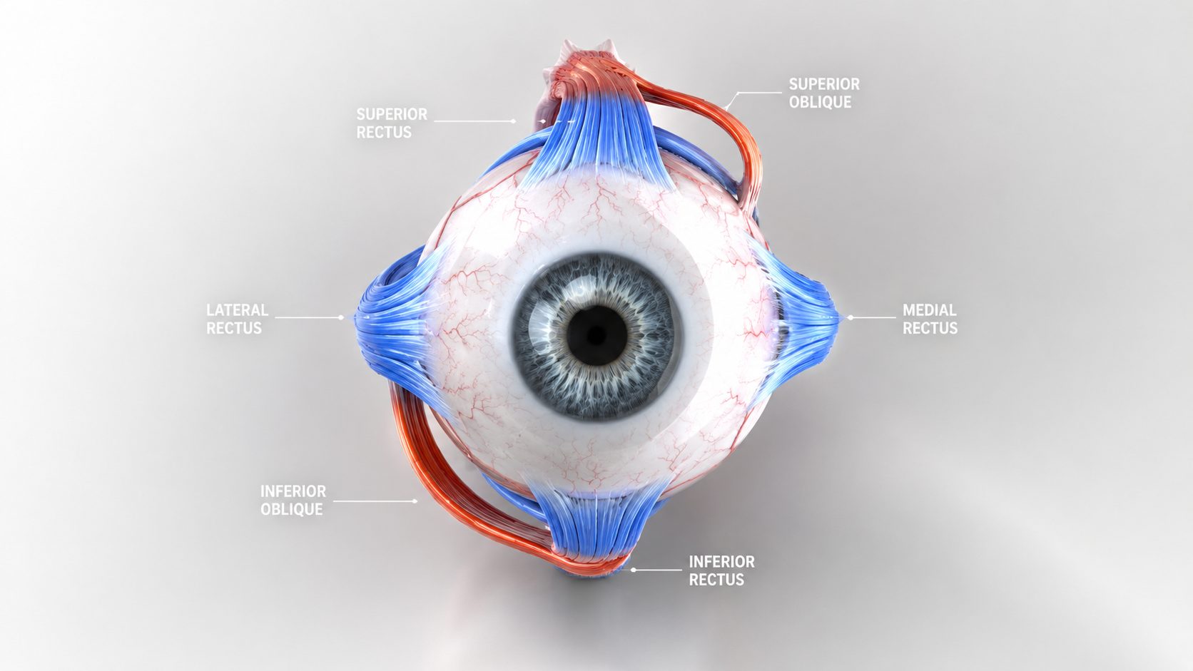
These are fast skeletal muscles, the same type that move your fingers. They generate two kinds of movement critical to photography:
- Saccades – (American-style: suh-KAHD / sak-AHD, British-style: sæk-AHD / sæk-KAHD) rapid, ballistic jumps between points of interest. When you scan a landscape for a composition, you’re firing hundreds of saccades per minute. Each one lands on a new fixation point in under 200 milliseconds.
- Smooth pursuit – the tracking movement that follows a moving subject. This only works for objects moving at moderate speed. When subjects move fast, the brain switches back to saccades to keep up.
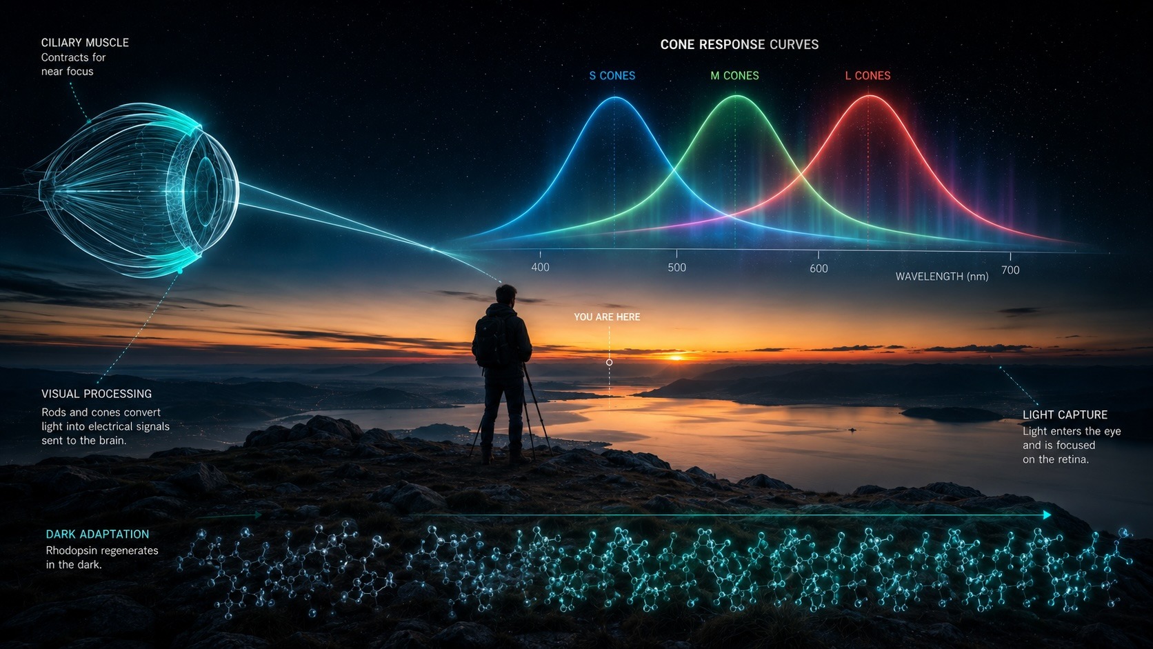
The focusing system. Inside the eye, the ciliary muscle forms a ring around the lens. When it contracts, it releases tension on the suspensory fibers (zonules) holding the lens flat. The lens rounds out, increasing its power to focus on close subjects. When the ciliary muscle relaxes, the zonules pull the lens flat again for distance. This is accommodation – the eye’s autofocus.
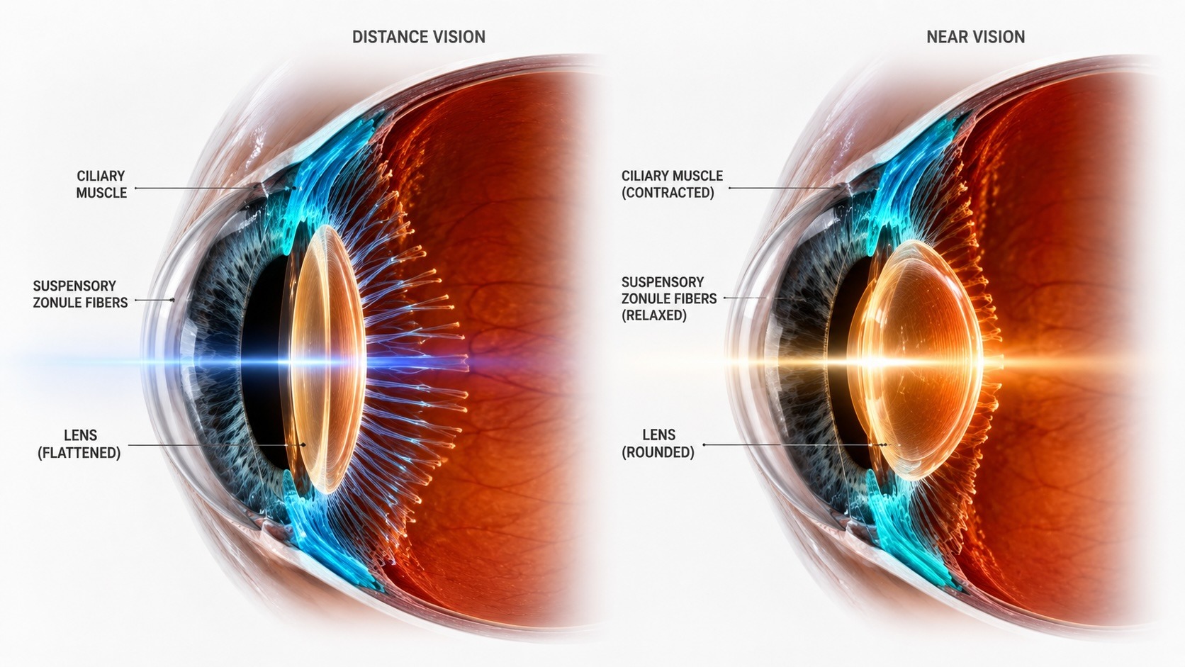
Here’s what makes this remarkable: the ciliary muscle is smooth muscle, the same type that runs your digestive system. It operates below conscious control. You don’t focus your eyes any more than you digest your lunch. The brain handles it automatically, thousands of times per day.
And like any muscle used constantly, it fatigues.
The Lens You Can’t Replace
The crystalline lens sits just behind the iris, suspended by those zonular fibers. In a young eye, it’s elastic and responsive – capable of shifting focus from infinity to a few inches in milliseconds.
This flexibility is accommodative amplitude, and it’s one of the most important optical properties your visual system has.
The lens is living tissue, but unusual living tissue. Its cells never shed. From the moment you’re born, new fiber cells wrap around the old ones, compressing the center of the lens over decades. By your early 40s, the lens core has stiffened enough that the ciliary muscle can no longer deform it fully. Near focus becomes harder. This is presbyopia, and it’s structural – a change in the physical material of the lens itself, not a muscle problem.
This distinction is important. Accommodative spasm (the ciliary muscle getting stuck in a contracted state from sustained near work) is functional and reversible. Presbyopia is structural and progressive. Exercises address accommodative, but they can’t reverse presbyopia.
What exercises can do – and the neuroscience here is exciting – is train the visual cortex to process lower-contrast, higher-frequency information more efficiently.
The brain learns to interpret the image the aging lens delivers. That’s a different mechanism, and it works.
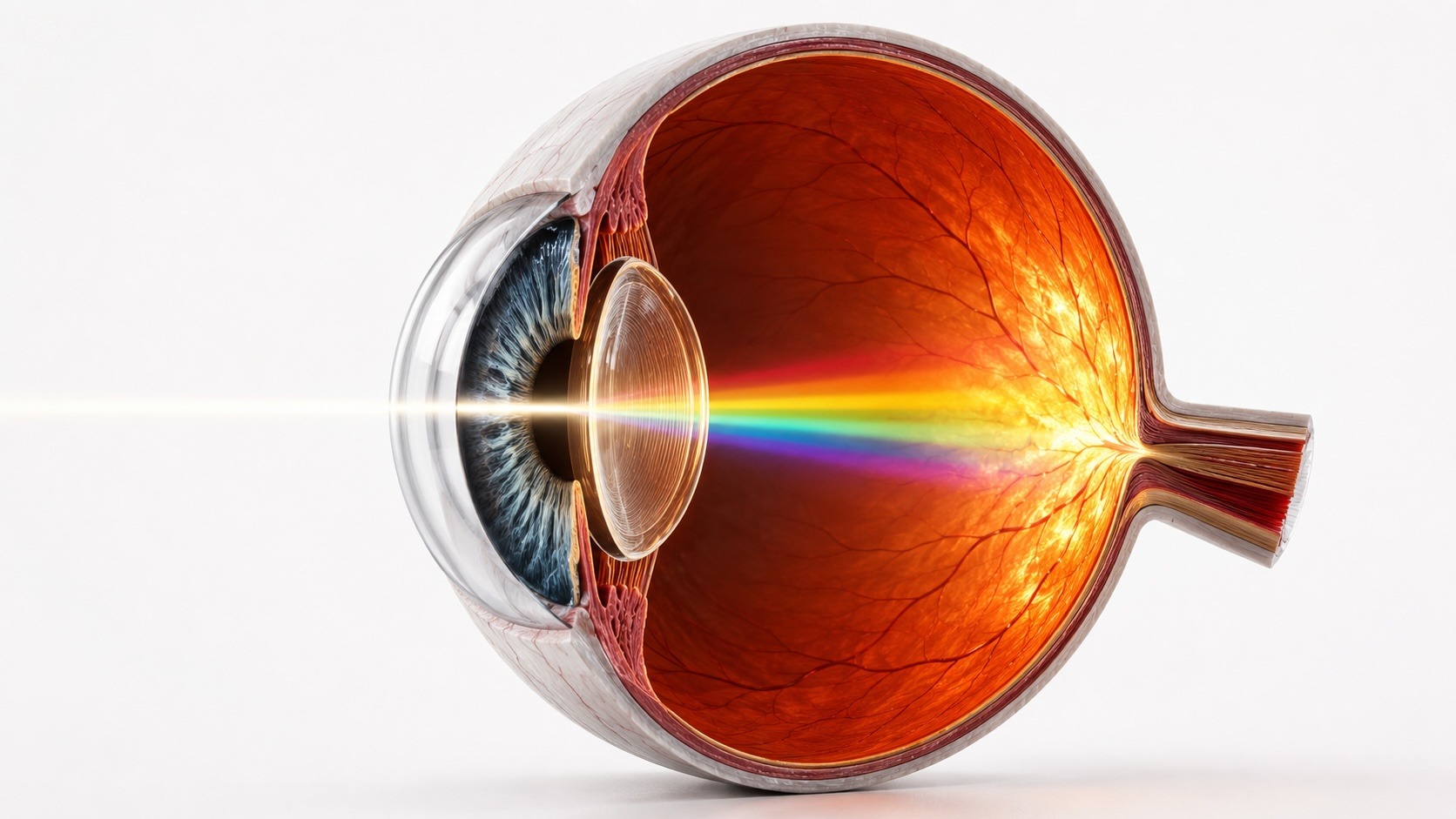
The Retina: Where Physics Becomes Perception
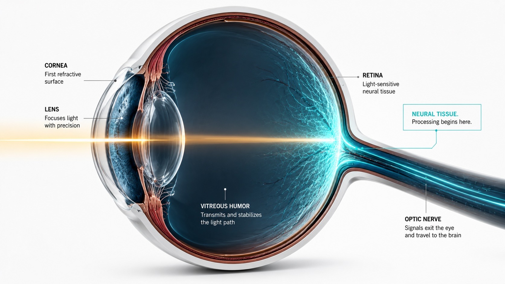
Light enters the eye, passes through the cornea and lens, travels through the vitreous humor, and lands on the retina – a thin sheet of neural tissue lining the back of the eye. The retina is, developmentally, a piece of the brain that migrated outward during embryonic development. It processes visual information before it even reaches the cortex.
The retina contains two types of photoreceptors, and understanding their differences is fundamental to photography in any given light.
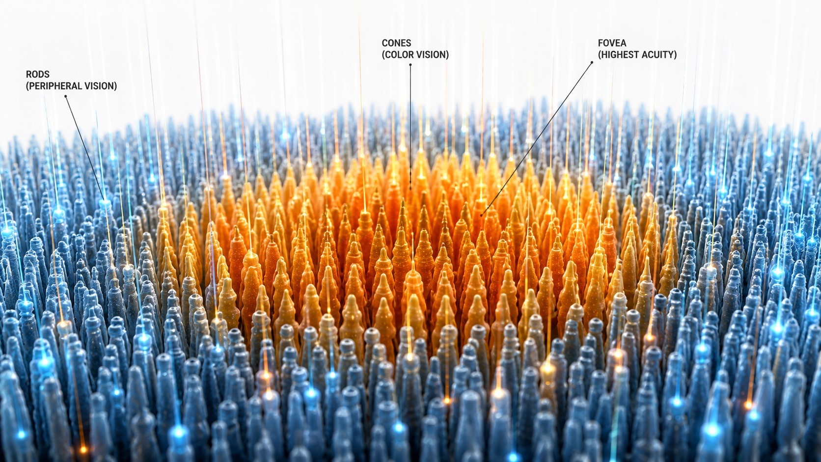
Cones. About 6 million of them, concentrated in the fovea – the small central pit at the center of the macula. The fovea is roughly 1.5mm in diameter. It handles color, fine detail, and bright-light vision. In the fovea, each cone connects to its own dedicated ganglion cell, giving you the highest possible spatial resolution at the center of your gaze. This is where you read. This is where you examine focus.
Rods. About 120 million of them, distributed across the peripheral retina with a gap at the fovea itself. The fovea has essentially zero rods. Rods don’t distinguish color. They’re achromatic, motion-sensitive, and extraordinarily sensitive to dim light – capable of responding to a single photon under ideal conditions. They pool their signals through shared ganglion cells, trading resolution for sensitivity.
The practical consequence: your central vision goes nearly blind in the dark. The fovea, so useful in daylight, becomes a liability at night. A faint star disappears when you look directly at it and reappears when you look slightly away – because you’ve shifted its image from rod-sparse fovea to rod-rich periphery.
Photographers who know this use it deliberately when framing night scenes and checking focus at the edge of darkness.
The Chemistry of Night Vision
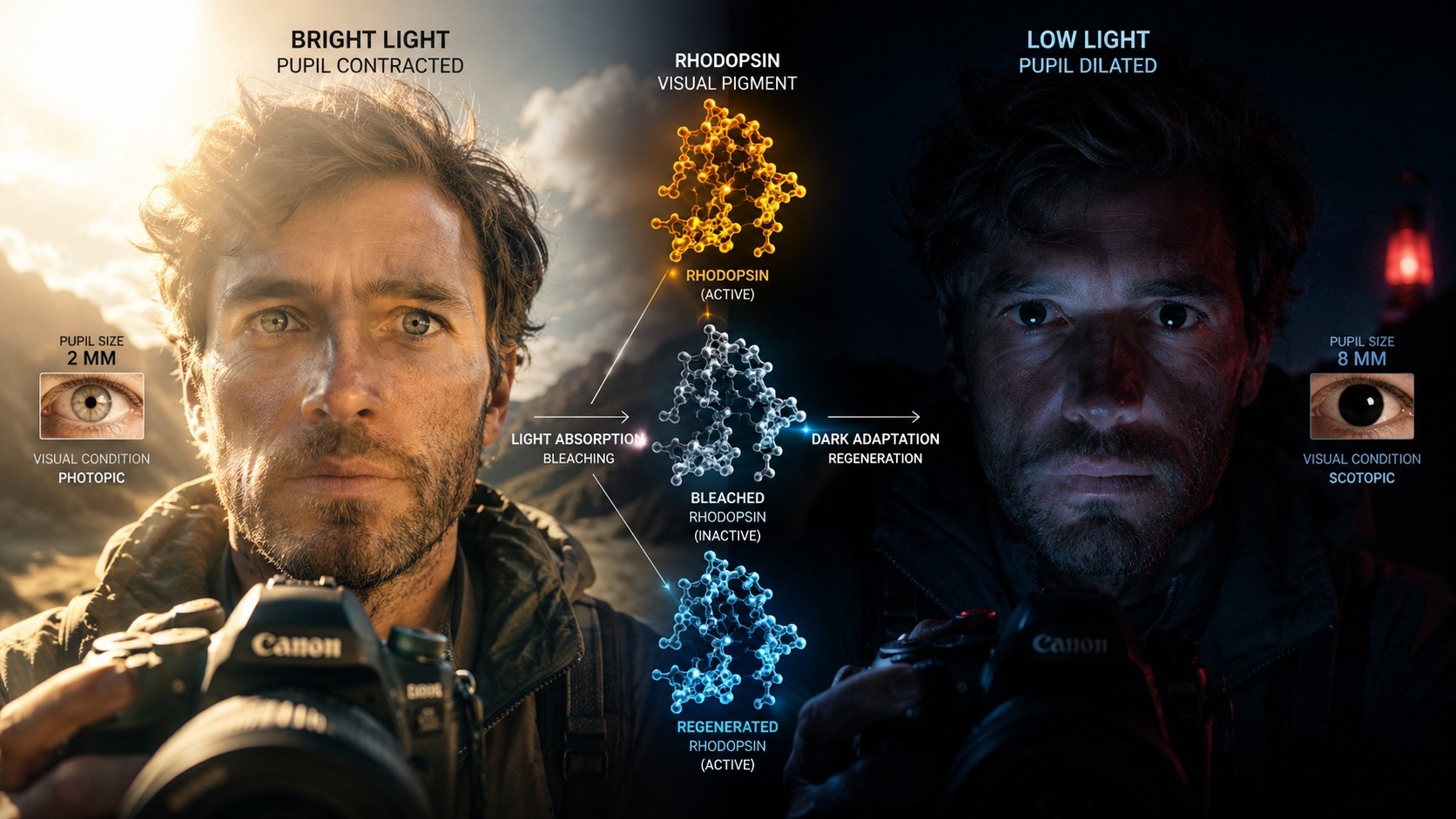
Rod photoreceptors run on a photopigment called rhodopsin – sometimes called visual purple. When a photon strikes rhodopsin, it triggers a molecular change that generates an electrical signal. That signal eventually reaches the brain as the experience of dim light.
The problem: rhodopsin bleaches when exposed to bright light. The molecule breaks down. It takes 20 to 40 minutes in darkness for rhodopsin to fully regenerate – and this regeneration is dark adaptation. Walk from a bright room into darkness, and your rods are largely offline for the first half hour.
White light resets this process almost instantly. A single glance at a phone screen in the dark costs you minutes of adaptation.
The solution photographers and astronomers have used for decades: red light. Rhodopsin’s peak sensitivity sits at around 500 nanometers – the blue-green range. Deep red light (above 620nm) barely registers on rods at all. A dim red headlamp or a camera display set to red mode preserves dark adaptation while still letting you check settings and navigate.
If you shoot landscapes, astrophotography, or any low-light work, arrive at your location 30 minutes before you need your full night vision. Stay off your phone. Use red light only. The images you get from a fully dark-adapted eye – both in terms of what you perceive in the scene and the compositional decisions you make – are different from what you’d get stumbling in from a brightly lit parking lot.
Color: Not What You Think
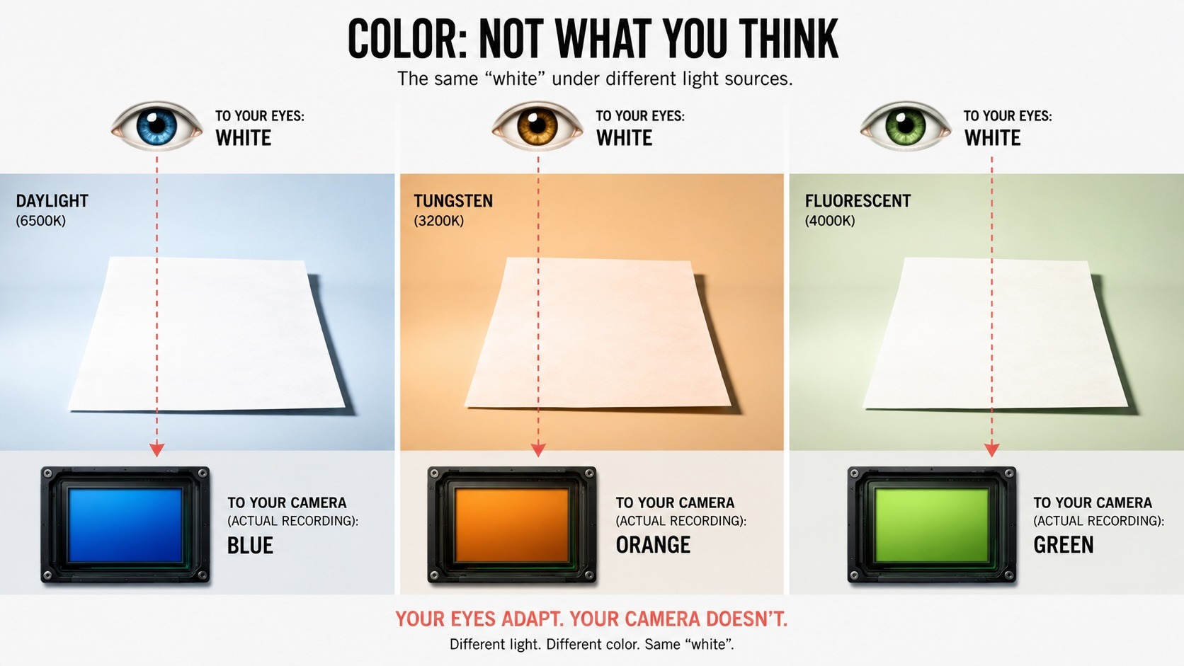
Color vision runs through three types of cones, each tuned to a different range of wavelengths – roughly corresponding to red, green, and blue (though the actual spectral curves are more complex and significantly overlapping). Color perception is the brain’s interpretation of the ratio of signals across these three populations, not a direct measurement of wavelength.
This means color is constructed. It’s an inference the brain makes from incomplete data.
White balance is a perfect example. Your visual system adapts so thoroughly to the color temperature of ambient light that you perceive a white sheet of paper as white whether it’s under daylight, tungsten, or fluorescent. The camera has no such adaptation – it records the actual spectrum. A warm-toned edit that looks neutral on a monitor calibrated for daylight looks radically different on a screen with a cooler white point.
The saturation spiral is what happens when you edit with fatigued eyes. As the ciliary muscle cramps and tear film degrades, light scattering increases in the eye itself. Perceived image contrast drops. The photographer compensates by pushing saturation and contrast in the edit. The next morning, with rested vision, the same image looks over-processed – colors punchy beyond recognition, shadows are crushed. The tools didn’t change. The visual system making the judgments did.
This is one of the most practical arguments for taking breaks during long editing sessions. The quality of your color decisions depends on the quality of your visual system in that moment.
Peripheral Vision: Your Compositional Radar
The periphery sees differently from the center – by design.
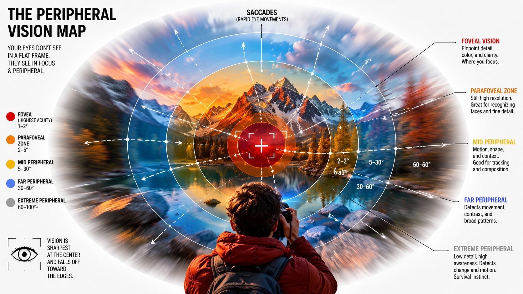
While the fovea delivers high-resolution color detail, the peripheral retina specializes in something else: scene gist.
In a single fixation, before any saccade, your peripheral vision captures the overall layout of a scene – the spatial relationships, the areas of brightness and shadow, the movement. This is the information that guides where you look next.
For photographers, peripheral awareness does something even more specific: it catches the unexpected. The gesture at the edge of the frame. The light breaking through clouds at the corner of the composition. The subject entering from frame left while you’re watching frame right.
Research on expert photographers and visual artists shows they develop more global scanning patterns – their eyes move through a scene in structured, efficient paths that build a complete spatial map rather than fixating on isolated details. This pattern develops with practice.
And… It’s trainable.
The peripheral retina also contains more motion-sensitive cells than the fovea. Your eye is wired to detect movement in the periphery and trigger a reflexive saccade toward it. This is ancient survival hardware built into the human eye, and it still serves photographers shooting unpredictable subjects.
Vision as Practice
Photography is, at its foundation, an act of seeing. The mechanics described here – the ciliary muscle pulling on zonule fibers, rhodopsin regenerating in the dark, cones comparing wavelength ratios to construct color – they’re the starting point of every image you’ve ever made.
Understanding them changes how you approach the craft. You arrive earlier for night shoots. You take breaks during editing sessions. You learn to trust the peripheral flash at the corner of your vision. You pay attention to how your perception shifts across a long day of shooting.
The eye is an active, adaptive system with its own rhythms, needs, and extraordinary capabilities. We should care for it accordingly.
In Article 2, we’ll look at what the evidence supports for training and protecting your vision – what works, what doesn’t, and what we as photographers should be doing daily.
Sources and References:
Eye Anatomy & Accommodation
- Physiology, Accommodation – StatPearls, NCBI Bookshelf, NIH https://www.ncbi.nlm.nih.gov/books/NBK542189/
- The Physiologic Mechanism of Accommodation – CRST Global https://crstodayeurope.com/articles/2014-apr/the-physiologic-mechanism-of-accommodation/
- Theories of Accommodation – ESCRS (European Society of Cataract & Refractive Surgeons) https://www.escrs.org/eurotimes/theories-of-accommodation
- Accommodation of the Eye to Different Focus Distance – HyperPhysics http://hyperphysics.phy-astr.gsu.edu/hbase/vision/accom.html
Digital Eye Strain & Tear Film
- Digital Eye Strain: A Comprehensive Review – PMC, NIH https://pmc.ncbi.nlm.nih.gov/articles/PMC9434525/
- Computer Vision Syndrome – American Optometric Association https://www.aoa.org/healthy-eyes/eye-and-vision-conditions/computer-vision-syndrome
Retinal Detachment & Red Flags
- Hollands H, et al. “Do findings on routine examination identify patients at high risk for primary open-angle glaucoma?” JAMA, 2009. (Meta-analysis: 14% of patients with acute-onset floaters/flashes have retinal tear)
https://pubmed.ncbi.nlm.nih.gov/19934426/
Viewfinder & Diopter
- Adjusting the Viewfinder to Your Eyesight – Exposure Therapy Photography Courses https://exposuretherapy.ca/photography-guide/adjusting-the-viewfinder-to-your-eyesight/
- Diopter Adjustment Errors: Hidden Risks – Chrosziel https://www.chrosziel.com/diopter-adjustment-errors-hidden-risks-for-accuracy-and-safety/
- Adjusting Your Diopter for Optimal Performance – Photofocus https://photofocus.com/photography/adjusting-your-diopter-for-optimal-performance/
Peripheral Vision & Scene Gist
- Eye Movement and Tracking in Sports – Specialty Vision https://specialty.vision/article/enhance-sports-performance-through-eye-movement-and-tracking/


Elevate Your Vision


Read this story and all the best stories on The Luminous Landscape
The author has made this story available to Luminous Landscape members only. Upgrade to get instant access to this story and other benefits available only to members.
Why choose us?
Luminous-Landscape is a membership site. Our website contains over 5300 articles on almost every topic, camera, lens and printer you can imagine. Our membership model is simple, just $2 a month ($24.00 USD a year). This $24 gains you access to a wealth of information including all our past and future video tutorials on such topics as Lightroom, Capture One, Printing, file management and dozens of interviews and travel videos.
- New Articles every few days
- All original content found nowhere else on the web
- No Pop Up Google Sense ads – Our advertisers are photo related
- Download/stream video to any device
- NEW videos monthly
- Top well-known photographer contributors
- Posts from industry leaders
- Speciality Photography Workshops
- Mobile device scalable
- Exclusive video interviews
- Special vendor offers for members
- Hands On Product reviews
- FREE – User Forum. One of the most read user forums on the internet
- Access to our community Buy and Sell pages; for members only.
You may also like